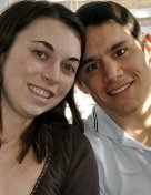
Leah is a college freshman who sings, dances, and plays the guitar. She is also a four-year survivor of an arteriovenous malformation (AVM).
It is rare for someone Leah’s age to have had an AVM, an abnormal tangle of blood vessels in the brain or spine that can cause devastating effects if it begins to bleed. Most likely the AVM had existed in Leah’s brain for many years, perhaps for most of her young life. She was fortunate to have received superb health care from doctors at Cincinnati Children’s Hospital Medical Center and from Dr. Andrew Ringer, a Mayfield Clinic neurosurgeon affiliated with The Neuroscience Institute at the University of Cincinnati and University Hospital.
In the fall of 2004 the AVM in Leah’s brain began to bleed, causing symptoms for the first time. Leah began experiencing headaches that, in retrospect, were more severe than ordinary headaches. “They came and went,” she recalls. “I had a pounding sensation in the back of head. I went to my doctor, and he thought I had a cold or virus. I took muscle relaxants for the pain, but it got worse. One day I had to sit out band practice because of the pain.”
Then, on October 6, 2004, the internal bleeding increased and Leah’s symptoms escalated.
“It was high school pajama day, which I was really looking forward to, Leah says. “I woke up with my head hurting really bad. My mom gave me some Tylenol and told me to lie down. I lay down for 10 minutes and woke up, and the room was spinning. My head was pounding. I threw up. I managed to yell for my mom and dad. I said you have to take me somewhere. The last thing I remember was my mom running to get her slippers. My dad carried me to the car and took me to the closest hospital.”
Hospital physicians ordered an MRI, which revealed that Leah was experiencing serious bleeding in the brain. The AVM, only one to two centimeters in diameter, was large enough to be life-threatening. Leah was then transported by helicopter to Cincinnati Children’s. She was in a coma for three days.
Doctors at Cincinnati Children’s stabilized Leah but decided that surgery on the AVM, which was located in the brainstem, should be postponed. The brainstem is a very sensitive area of brain, and the risk of coma or death as a complication of surgery is much higher than with other areas of brain The Cincinnati Children’s team wisely chose not to operate immediately after the hemorrhage occurred.
After spending three weeks in the hospital, Leah was able to go home. Her doctor told her he was handing her case over to Dr. Ringer, a Mayfield Clinic neurosurgeon who eventually would perform her surgery. Leah left with warm memories of Cincinnati Children’s. “I loved that hospital,” Leah says. “I love it to this day. Everyone was so nice there. Everyone complimented me. They would say, ‘You’re so pretty.’ When I left the hospital it was kind of a shock; I was out in the real world where everyone wasn’t as nice.”
Leah also faced difficult adjustments in her day-to-day life. It was critical that no sudden movement cause the AVM to begin bleeding again. “When I was released from the hospital, they gave me information about what I could and couldn’t do,” Leah says. “I wasn’t allowed to do anything that would cause my brain to be active. I couldn’t do a cartwheel, play my clarinet, or work out. It was torture. It was horrible.
"In April 2005, Leah was ready for surgery. Dr. Ringer, who directs the endovascular neurosurgery program at the University of Cincinnati Academic Health Center, operated on Leah at University Hospital. Leah’s grandmothers prayed over Dr. Ringer’s hands before the surgery began. Then, over a period of seven hours, Dr. Ringer performed an innovative, minimally invasive procedure to repair the malformation.
“We used an endovascular approach, entering the artery in the groin and then navigating the arteries to the basilar artery at the brainstem,” Dr. Ringer says. “We used a small catheter that is slightly less than 1 millimeter in diameter at its tip to navigate the small arteries that fed the AVM.”
Once Dr. Ringer had reached the AVM with the tiny catheter, he used a technique called embolization to block the blood flow through each of the pathological arteries that was feeding the AVM. Deprived of blood flow, the AVM could no longer bleed. “Traditionally this technique has been used to block some, but not all the arteries to an AVM in preparation for surgery or radiation,” Dr. Ringer says. “We felt that neither of these were good options for her, so we spent a lot more time and effort trying to block every one of the abnormal arteries. Fortunately, it worked.”
Dr. Ringer told Leah she wouldn’t have to worry about the AVM again – that she could return to dancing, running, singing, and playing the clarinet with her high school band. She was happy to oblige. Four years later she is taking classes, enjoying her friends, playing the guitar, and eyeing a possible future in the field of nutrition, health, or music. Says Leah: “I am so blessed to have my life back.”
 RSS Feed